

What Is Endometriosis Mapping?
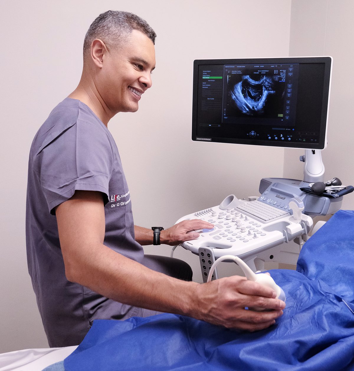
Endometriosis mapping is a specialised imaging assessment that charts the position and depth of endometriosis throughout the pelvis and, where relevant, beyond it. It is performed mainly with a transvaginal ultrasound in both 2D and 3D, following a structured protocol developed by the International Deep Endometriosis Analysis (IDEA) group, an international panel of specialists in endometriosis imaging. Where needed, MRI adds information about areas the ultrasound cannot reach.
A routine gynaecological scan focuses on the uterus and ovaries. Mapping goes considerably further, deliberately examining the ligaments that support the uterus, the wall of the bowel, the bladder and the ureters, as well as the space between the uterus and rectum. This is where deep endometriosis tends to hide, and it is why a woman can be told her scan is normal while still living with significant disease.
Why Is Endometriosis Mapping So Important?
For many years, laparoscopy was regarded as the only way to diagnose endometriosis. That has changed. Current international guidance, including the 2022 guideline from the European Society of Human Reproduction and Embryology (ESHRE), recognises imaging as the first-line diagnostic tool, especially for more severe disease, with laparoscopy reserved for women whose scans are negative or whose symptoms continue despite medical therapy. You can read more about this shift on the diagnosing endometriosis page.
The real value of mapping, though, lies in what happens next. Knowing where your endometriosis is, and how deeply it has grown, before any decision is made means that:
- Treatment can be matched to the disease you actually have, whether that points towards medical therapy or surgery
- Surgery, when it is needed, can be planned in full beforehand, which reduces the likelihood of an incomplete operation and a second procedure later
- A colorectal surgeon or urologist can be arranged ahead of time when the bowel or urinary tract is involved
- You can have an honest conversation about what surgery would involve for you, including recovery time and any implications for fertility, before the day rather than after it
What Does Endometriosis Mapping Look For?
Each part of the scan is aimed at a specific question about your endometriosis. The main findings Dr Groenewald looks for include the following.
Ovarian Endometriomas
Endometriomas are cysts on the ovary filled with old blood, sometimes called chocolate cysts. They are usually straightforward to see on ultrasound. Their size, and whether they affect one or both ovaries, matter a great deal for treatment decisions and for protecting your future fertility.
Deep Infiltrating Endometriosis
Deep infiltrating endometriosis grows more than five millimetres beneath the surface of the tissue it affects. It forms firm nodules, most commonly in the uterosacral ligaments behind the uterus and in the rectovaginal septum, the tissue between the vagina and rectum. It can also grow into the walls of nearby organs. These nodules are often the source of deep pain during sex and with bowel movements. You can read more about how deep endometriosis develops.
Bowel Endometriosis
When endometriosis involves the bowel, it most often affects the rectum and sigmoid colon. Mapping records the size of each nodule and how deeply it has grown into the layers of the bowel wall. It also measures how far the nodule sits from the anal opening. These measurements are exactly what a colorectal surgeon needs to decide, together with Dr Groenewald, whether a nodule can be shaved from the bowel surface or whether a small section of bowel needs to be removed.
Urinary Tract Endometriosis
Endometriosis can form nodules within the bladder wall, which may cause pain or urgency when passing urine around your period. It can also surround the ureters, the narrow tubes that carry urine from the kidneys to the bladder. Ureteric endometriosis often causes no symptoms at all, yet over time it can narrow the ureter and cause the kidney to swell. For this reason, mapping includes a scan of both kidneys, so that any involvement of the urinary tract is known and a urologist can be included in planning.
Diaphragmatic Endometriosis
Occasionally, endometriosis is found outside the pelvis on the diaphragm, the muscle beneath the lungs. It typically causes pain in the right shoulder tip or chest that follows your cycle. A pelvic ultrasound cannot see the diaphragm, so these symptoms are noted carefully during your consultation. MRI may be helpful, and where diaphragmatic disease is suspected, the surgical plan includes a thorough inspection of the upper abdomen.
Adenomyosis and Adhesions
Adenomyosis, in which endometrial-like tissue grows into the muscle wall of the uterus, often occurs alongside endometriosis and can cause heavy, painful periods of its own. 3D ultrasound is particularly useful for assessing it. Mapping also checks whether organs have become stuck together by adhesions, which affects both the complexity of surgery and the likelihood of pain.
How Is Endometriosis Mapping Performed?
2D and 3D Transvaginal Ultrasound
The scan is performed with a slim ultrasound probe placed in the vagina, which allows a close, clear view of the pelvic organs. Following the IDEA approach, it works through four steps in a set order:
- The uterus and ovaries are assessed, including for adenomyosis and endometriomas
- “Soft markers” are checked, such as tenderness at specific points and how freely the ovaries move
- The sliding sign is tested, a gentle check of whether the uterus and bowel glide over each other or are held together by adhesions
- The spaces in front of and behind the uterus are searched carefully for deep endometriosis nodules, including those in the bowel and bladder
3D ultrasound captures a volume of information rather than a single flat image. This allows the uterus to be viewed from angles that 2D scanning cannot show, which helps with assessing adenomyosis and the shape of the uterus, and with measuring nodules accurately. A short abdominal scan of the kidneys completes the assessment.
When Is MRI Used?
MRI complements ultrasound rather than replacing it. It may be recommended when endometriosis appears to extend higher into the bowel than the ultrasound probe can reach, when disease involves the pelvic sidewall or nerves, or when diaphragmatic endometriosis is suspected. It is also useful when planning complex or repeat surgery, and when a transvaginal scan is not suitable for you. Where MRI is needed, it is arranged through a radiology practice, and Dr Groenewald reviews the images alongside the ultrasound findings.
Endometriosis Mapping in Cape Town
Dr Groenewald performs endometriosis mapping at his practice at Life Kingsbury Medical Suites in Claremont, for patients across Cape Town and further afield. If you have been told your scan was normal, but your symptoms say otherwise, or if you have been advised to have surgery and want to understand the full extent of your disease first, a mapping assessment is a practical next step.